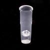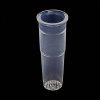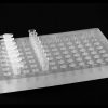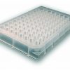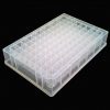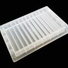
The mPrep System™ streamlines Transmission Electron Microscopy (TEM) sample preparation using a capsule based approach. Use mPrep/s capsules to fix, orient, embed, and section specimens. Use mPrep/g capsules to stain or immuno-label TEM grids.
The mPrep System efficiently produces quality results from every sample. Imagine this in your lab.
- As few as two human touches from microtome to microscope – reduces damage and loss
- Grids and capsules labeled for easy tracking from start to storage
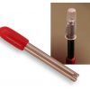
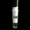
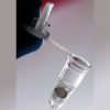
- Capsules attach to common lab pipettors for controlled reagent timing and minimal reagent consumption
- Parallel processing
- Stain from one to dozens of grids simultaneously using multi-channel pipettors
- Automate using the mPrep-ASP-1000 Automated Specimen Processor
- Identical reagent timing
- Reduced tedium and labor costs
The mPrep System is designed for versatility. Our customers continue to develop new applications. Explore some TEM applications below.